CARDIOVASCULAR SYSTEM

CARDIOVASCULAR SYSTEM
ORGANS ASSOCIATED :


STRUCTURE OF HEART :
HEART is a hollow muscular vascular organ that weighs around 250 gm in females and 350 gm in males, depending upon the body size, Age and body weight .
It is a cone shaped organ .
It is located in Mediastinum between the lungs .
its 2/3 rd mass lies to the left and 1/3 rd mass lies to the right of Mediastinum .
Its apex is located below and its base is located above .
Its apex lies at the level of 5th inter-coastal space .
Base of the heart lies at the level of second inter-coastal muscles.
Measurement of heart are, its length is 12 centimetre, width is 9 centimetre and height is 6 centimetre .
SUPERIOR : Greater blood vessels such as Pulmonary Artery and Aorta .
INFERIOR : Diaphragm , Liver, Stomach, Small intestine , Large intestine.
POSTERIOR : Aorta, Pulmonary veins.
ANTERIOR : Sternum, Inter-coastal muscles , Lungs and Ribs .
Heart has three Layers.
(1) PERICARDIUM
(2) MYOCARDIUM
(3) ENDOCARDIUM
(1) PERICARDUIM :
Pericardium is the outermost layer of heart.
It is further divided into two layers.
Parietal pericardium and Visceral pericardium.
There is a space between Parietal and Visceral pericardium, known as Pericardial cavity.
Fluid is present in the Peri-Cardial space about 30 ML. fluid is present in the pericardial space .
(2) MYOCARDIUM :
Myocardium is the middle layer of heart.
it is made up of specialized cardiac muscle cells which are responsible for contraction of heart .
(3) ENDOCARDIUM :
It is the innermost layer that lines the heart structure, chambers and the Valves of the heart.
Blood vessels that arise from heart are in direct contact with the Endocardium.
It provides smoothness to heart and blood vessels .
CHAMBERS OF HEART :
Heart is divided into four chambers.
The upper two chambers are called right and left atria.
The lower two chambers are called right and left ventricles.
The right and left atrium are separated by a partition called Inter-atrial septum.
The right and left ventricles are separated by intraventricular septum .
RIGHT ATRIUM STRUCTURE : Thinner Walls.
LEFT ATRIUM STRUCTURE : Slightly Thicker Walls as compared to Right Atrium.
RIGHT VENTRICLE : Thicker in size.
LEFT VENTRICLE : Most Thickest in size.
STRUCTURE OF HEART :
VALVES OF HEART :
(1) ATRIOVENTRICULAR VALVES :
These Valves line between Atrium and Ventricles.
TRICUSPID VALVE :
It lie between right Atrium and right Ventricle . It has three cusps.
BICUSPID VALVE :
It is also known as Mitral Valve . It lie between left atrium and left ventricle.
it has two cusps
(2) SEMILUNAR VALVES :
Semi-lunar valves are size of half moon shape.
(1) PULMONARY VALVE :
Pulmonary Valve is a Semi-lunar valve that lies between Right Ventricle and Pulmonary Artery.
(2) AORTIC VALVE :
Aortic Valve is also a Semi-lunar valve, that lies between left Ventricle and Aorta.
ARTERIAL BLOOD SUPPLY TO HEART :
Right Coronary Artery supplies the right side of Heart.
Left Coronary Artery supplies the left side of Heart.
Descending Artery supply the left Ventricle and Ventricular Septum.
Heart receives 5% of Blood from total Cardiac output.
VENOUS SUPPLY :
Coronary Sinus Drain the whole De-oxygenated Blood from the Heart into the Right Atrium .
PHYSIOLOGY OF HEART :
Heart is a pumping organ.
When the myocardium contracts heart start beating, and beats throughout the life beginning at 16 weeks of gestation, Heart start beating with each beat heart pumps blood to the two major arteries.
Heart performs two major circulation .
(1) PULMONARY CIRCULATION :
Superior and inferior Vena Cava collects de-oxygenated blood from the whole body and supply it to the right side of the heart .
From Superior and Inferior Vena Cava blood moves to the Right Atrium.
From Right Atrium blood moves to the Right Ventricle via Tricuspid valve.
From Right Ventricle blood moves to the Pulmonary Trunk via Pulmonary valve.
From Pulmonary Trunk the blood moves to the Right and the Left Pulmonary Artery.
From Right Pulmonary Artery , The Blood moves to the right lung.
From the left Pulmonary Artery it moves to the left lung .
In the right and the left lung exchange of gases takes place at alveoli space .
Right and Left Pulmonary Artery is further divided into arterioles and subdivided into capillaries .
The capillaries goes deep inside the tissues of the lungs .
Alveoli exist at the tissue level in the lungs.
Alveoli are tiny Air Sacs at the end of the Bronchioles in the Lungs.
Alveoli and Capillaries have thin semi permeable membrane which allows exchange of gases between blood and Alveoli.
Diffusion takes place inside the lungs and the carbon dioxide moves from blood to the alveoli and oxygen moves from alveoli to the capillaries .
Oxygenation of Blood takes place inside the Lungs.
Release of Carbon dioxide as a waste product from the lungs takes place known as Expiration.
FLOWCHART
Veins collect Blood from Whole body, Upper ,Central and
Lower body and Drains it into 2 Major Veins, SUPERIOR AND
INFERIOR VENA CAVA
↓
RIGHT ATRIUM
↓ Via Tricuspid valve
RIGHT VENTRICLE
↓ Via Pulmonary Valve
PULMONARY ARTERY
↓
Right and Left lung(Exchange of gases takes place,blood gets Oxygenated in Lungs)
(2) SYSTEMATIC CIRCULATION :
The pulmonary veins carries oxygenated blood from right and left lung to the heart .
Blood moves from Pulmonary veins into the Left Atrium . From Left Atrium blood moves into the Left Ventricle via mitral valve.
From Left ventricle blood moves to the Aorta via Aortic
valve.
From Aorta blood moves to the whole body parts. Aorta arise into three main branches Arch of Aorta, Ascending Aorta ,and Descending Aorta.
All these branches further divide and subdivide supplying the whole body organs and tissues .
FLOWCHART
PULMONARY VEINS
↓
LEFT ATRIUM
↓ (via Mitral Valve )
LEFT VENTRICLE
↓ ( Via Aortic Valve )
AORTA
↓ ( Via its branches )
TO THE FULL BODY
CONDUCTING SYSTEM OF HEART :
It involves the Electrical Activity of the Heart.
Four structures are responsible for Heart Conduction.
(1) SA NODE (SINOATRIAL NODE ) :
SA Node is located on superior side in Right Atrium near the opening of Superior Vena cava.
Group of neuron cells are present in the sinoatrial node.
SA Node initiate the nerve impulses signal. SA node is called pacemaker of the heart .
(2) ATRIOVENTRICULAR NODE :
Cardiac muscle tissue is present in the AV node . Atrioventricular node lies in the right side between the atrium and interatrial septum .
(3) ATRIOVENTRICULAR BUNDLE :
AV bundle arises in the AV node and extend to the sides of interventricular septum.
AV bundle further extent to the Perkinjee fibre.
(4) PURKINJEE FIBRE :
Perkinjee fibre are made up of cardiac muscles responsible for contraction of Heart.
Perkinjee fibre are responsible for transmitting electrical impulses from the Atrio- ventricular node to ventricles of Heart .
NERVE SUPPLY TO HEART :
Sympathetic and parasympathetic nerve fibre form a cardiac plexus in the heart .
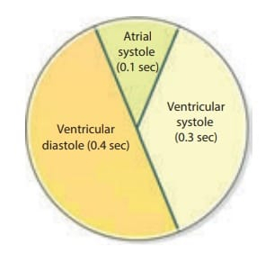
CARDIAC CYCLE :
Cardiac cycle involves a series of events that takes place in the circulation of blood, while heart is pumping. It involves Systole and Diastole phase .
(1) ATRIAL SYSTOLE :
Contraction of Atria.
Duration - 0.1 second
(2) VENTRICULAR SYSTOLE :
Contraction of Ventricles Duration - 0.3 second
(3) COMPLETE CARDIAC DIASTOLE :
It involves both relaxation of both Atria and ventricles . Duration - 0.4 second
TOTAL DURATION OF CARDIAC CYCLE : 0.8 SECOND
Heart is a pumping organ it beats throughout the life starting from intrauterine period.
Atrium and ventricles put their respective blood into the Pulmonary Artery and Aorta by their contraction.
Contraction of Atria is shorter 0.1 second, ventricles contact for a longer duration that is 0.3 second and with a greater force.
Sinoatrial Node triggers a wave of contraction and the blood moves from Atria to ventricles that is known as atrial Systole , that is 0.1 second.
Then the waves of contraction reaches the AV Node and contraction reaches to ventricular muscle fibre via AV Bundle, Bundle of His and Perkinjee fibre.
Then Ventricular systole takes place, ventricles pump their blood into the Pulmonary Artery and Aorta .
The Valves b/w Atria and Ventricles and the Semi-lunar valves open and close depending upon pressure of Blood.
When pressure increases the Valve gets Open,and Closes when the Pressure of Blood Reduces
HEART SOUNDS :
Two Heart sounds are produced during Cardiac Cycle.
S1 and S2
S1 : S1 Sound is produced due to closure of
Atrioventricular Valves.
S2 : S2 Sound is produced due to closure of Semi-lunar Valves.
S1 Sound is known as Lub sound.
S2 Sound is known as Dub sound.
ECG (ELECTRO CARDIO GRAPH)
ECG Shows the Electrical activity of Heart in the form of graph.
Normal ECG Consists of P Wave , QRS Complex and T Wave.
P Wave Represent the Depolarization of Atria.
QRS Complex Represent Depolarization of Ventricles , and Re-polarization of Atria.
T Wave Represent Re-polarization of Ventricles.
Depolarization means Contraction .
Re-polarization means Relaxation .
Sinus Rhythm of Heart is 60-100 Beats/Min.
Tachycardia - Heart Rate more than 100 Beats/Min.
Bradycardia - Heart Rate less than 60 Beats/Min.
CARDIAC OUTPUT :
The total Amount of Blood Pumped by Heart in a minute is known as Cardiac Output.
CALCULATION OF CARDIAC OUTPUT : CARDIAC OUTPUT = STROKE VOLUME * HEART RATE STROKE VOLUME :
Amount of Blood Ejected by both Ventricles in each Beat / Contraction is known as Stroke Volume . It is
About 70 ML
Cardiac Output of a human being lies B/W 4 To 6 Litre of Blood per minute.
BLOOD PRESSURE :
It is the Pressure exerted by the Blood on the walls of Blood Vessels.
(1) SYSTOLIC PRESSURE :
It is the pressure produced in the Aorta and Pulmonary Artery , during the Ventricular Systole.
It Range B/W - 100 TO 140 mmHG Average Systolic pressure is 120 mmHG
(2) DIASTOLIC PRESSURE :
Period of Diastolic phase, when the Heart /Ventricles are in resting phase, the pressure inside the Blood Vessels in that phase is less is known as Diastolic Pressure.
Range of Diastolic Pressure - 60 to 90 mmHG Average Diastolic Pressure is 80 mmHG
(3) PULSE PRESSURE :
It is the difference between the Systolic and Diastolic Blood Pressure.
FACTORS AFFECTING BLOOD PRESSURE :
(1) BLOOD VOLUME :
Normal range of blood volume is 4 TO 6 Litre in human body.
Increase/decrease in the level of volume of blood causes change in Blood Pressure.
HYPERTENSION :When pressure raises above 140/90 mmHG
HYPOTENSION : When pressure decrease below
100/60 mmHG
(2) CARDIAC OUTPUT :
Increase/ decrease in cardiac output causes change in blood pressure .
(3) BLOOD VISCOSITY :
Viscosity means Resistance to flow. A thick Blood causes more pressure on the walls of Blood Vessels.
More the Viscosity of Blood more will be the Blood Pressure.
(4) VENOUS RETURN :
The Amount of Blood returned by the Superior and Inferior Vena cava, play an important role in Cardiac Output.
(5) ELASTICITY OF BLOOD VESSELS :
With ageing the Vessels Elasticity Reduces, and the Blood Pressure Arises.
REGULATION OF BLOOD PRESSURE :
BARORECEPTORS :
These are the Specialized Nerve cells located in the Aortic Arch and Carotid Sinus.
When Blood Pressure is Raised in the Blood vessels these cells respond by sending Signal to Cardiovascular Centre located in Brain.
Cardiovascular Centre respond by Increasing Para sympathetic stimulation which decrease the Heart Rate, Cardiac Output is reduced, Hence Blood Pressure is Reduced.
In case of Low Blood Pressure in these Blood Vessels , these cells send Signal to Cardiovascular Centre in the Brain.
The Brain Respond by Stimulation of Sympathetic Nerve Supply which cause Vasoconstriction.
PULSE RATE : DEFINITION :
It is defined as Alternate Expansion and Recoil of an Artery due to Contraction of Heart and Ejection of Blood in already Full Aorta.
OR
Number of Time , Heart Beats within A minute is called as Pulse Rate.
CHARACTERISTICS OF PULSE RATE :
(1) RATE : AT Which Heart Beats Per Minute.
(2) RHYTHM :It Refers to the Regularity of Pulse weather Regular or irregular.
(3) VOLUME : It Refers to Strength of Expansion and
Contraction.
A strong Pulse indicate Adequate Blood Flow. A Weak Pulse Indicate decreased Cardiac Output and State of Hypovolemia.
(4) TENSION :
A LOW Tension Pulse means the vessel feels soft or Impalable b/w the Beats.
A High tension Pulse means the Vessels feel Rigid between Beats.
AORTA AND ITS BRANCHES :
Aorta is the largest Artery in the Human Body.
Aorta is Divided into 3 Main branches.
(1) Ascending Aorta
(2) Arch of Aorta
(3) Descending Aorta
(1) ASCENDING AORTA :
It has 2 main branches.
Right Coronary Artery Left Coronary Artery
(2) Arch of Aorta :
(1) Brachiocephalic Artery
(2) Left Common Carotid Artery
(3) Left Subclavian Artery
(3)Descending Aorta :
Descending Aorta is down with Thoracic and Abdominal Aorta and their further branches.
(1) Thoracic Aorta :
Branches of Thoracic Aorta : Brachial Arteries
Oesophageal Arteries Intercoastal Arteries
(2) Abdominal Aorta :
Branches of Abdominal Aorta :
Renal Artery
Left Gastric Artery
Testicular Artery
Inferior Phrenic Artery
Lumbar Artery
Splenic Artery
Common Hepatic Artery
(3) BLOOD CIRCULATION TO LOWER LIMBS :
Common Illac Artery Plantar Artery
BLOOD CIRCULATION TO HEAD AND NECK :
It comes under CIRCLE OF WILLIS
Common Carotid Artery and Vertebral Arteries supply blood to Head and Neck with their further branches.
(1) External Carotid Artery
(2) Internal Carotid Artery
CIRCLE OF WILLIS (HEAD SUPPLY ) :
2 Anterior Cerebral Arteries
2 Internal Carotid Arteries
1 Anterior Communicating Artery
2 Posterior Communicating Arteries
2 Cerebral Arteries
1 Basiliar Artery
ARCHOFAORTA
↓ ↓ ↓
BRACHIOCEPHALIC LEFT SUBCLAVIAN LEFT
TRUNK ARTERY COMMON CAROTID
↓ ↓ ARTERY
(1)RIGHT SUBCLAVIAN (1) LEFT VERTEBRAL
ARTERY ARTERY
↓ (2)LEFT AXILLARY ARTERY
(a) RIGHT VERTEBRALARTERY
(b) RIGHT AXILLARYARTERY
↓
RIGHT BRACHIAL ARTERY
↓
(a) RIGHT RADIALARTERY
(b) RIGHT ULNARARTERY
↓
RIGHT SUPERFICIAL AND
DEEP PALMER ARCHES
↓
RIGHT DIGITALS
↓
(1) LEFT RADIAL ARTERY
(2) LEFT ULNAR
ARTERY
↓
LEFT SUPERFICIAL
AND DEEP PALMER ARCHES → LEFT
DIGITALS
(2)RIGHT COMMON
CEREBRAL ARTERY
↓
(1)RIGHT EXTERNAL
CAROTID ARTERY
(2)RIGHT INTERNAL
CAROTID ARTERY